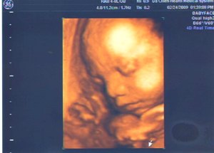
4D立體超音波不但解析度清晰細緻,而且可以「同步即時」的觀看胎兒動態的立體影像,使肚子裡胎兒的一舉一動,完整呈現,讓媽咪對寶寶的印象不再只是虛擬的想像,而是最真實的影像記錄。
 「4D立體超音波」是目前產科最進步的超音波,最新的立體4D超音波以極短的時差來呈現即時的胎兒動態影像,也是醫學診斷科技的一大進步。它除了能夠讓我們看到3D立體影像外,還能看到「即時」的胎兒動態影像。我們在掃描過程中可以調整X、Y、Z軸,如此更能清楚的看到胎兒上下、前後、左右的相關位置。另外準爸爸媽媽們可以利用「4D立體超音波」看到胎兒在子宮內伸伸手、踢踢腳、吸大拇指、打哈欠…等臉部表情,放鬆心情,舒緩即將為人父母的緊張情緒。
在一般產檢時所做的超音波只能看胎兒大小、胎位、胎盤位置、羊水量及胎兒較大的畸型,稱為初步的超音波檢查。但是對於有特殊病史的產婦,或是產檢發現有問題時,可以進一步做高層次超音波檢查。通常高層次超音波是以解析度精良的2D超音波執行,必要時用3D、4D或杜卜勒超音波來輔助診斷。此項檢查需由經過特別訓練的醫師專家來執行,檢視許多胎兒更細部的異常,如心臟、腦部、內臟…結構異常等。建議於妊娠20至24週(即懷孕5~6個月)執行高層次超音波最為恰當。
有以下情況的產婦,建議進一步做高層次超音波檢查:
1.常規檢查或母血篩檢值異常,懷疑有胎兒畸型者
2.早期妊娠有接觸不明藥物或不明感染者
3.有胎兒生長遲滯現象者
4.羊水量過多或過少者
5.前胎有生產畸型胎兒者
6.懷孕合併內科疾病者,如糖尿病、自體免疫疾病等。
There are differences in the types of ultrasounds done in pregnancy. The old standard was a 2D or 2 dimensional image. In recent years this has been replaced with the new 3D or three dimensional images and now 4D.
2D ultrasound give you outlines and flat looking images, but can be used to see the internal organs of the baby. This is helpful in diagnosing heart defects, issues with the kidneys and other internal issues. The 3D images are used to show you three dimensional external images which may be helpful in diagnosing issues such as a cleft lip. What 4D ultrasound brings to the table is that as the image is continuously updated, it becomes a moving image, like a movie.
Elective ultrasounds are a great way for families to bond with their unborn baby. Being well-prepared can help you have the best experience possible when getting that first amazing glimpse of baby!
What are 3D/4D ultrasounds, and why is everyone
As
an expectant mother, you’re excited to see your baby at every stage,
from the first little dot on the monitor to the sweet kicking baby ready
to enter this world. Every ultrasound is a moment to treasure.
Is 3D/4D ultrasound the best way to learn the gender of my baby?
It may surprise you to learn that 2D ultrasound is the best way to determine or verify a baby’s gender. 3D ultrasound can, at times, be a bit ambiguous when it comes to gender images. Other times, it’s quite obvious. However, the best way to make sure of an accurate gender determination is through 2D ultrasound.
 「4D立體超音波」是目前產科最進步的超音波,最新的立體4D超音波以極短的時差來呈現即時的胎兒動態影像,也是醫學診斷科技的一大進步。它除了能夠讓我們看到3D立體影像外,還能看到「即時」的胎兒動態影像。我們在掃描過程中可以調整X、Y、Z軸,如此更能清楚的看到胎兒上下、前後、左右的相關位置。另外準爸爸媽媽們可以利用「4D立體超音波」看到胎兒在子宮內伸伸手、踢踢腳、吸大拇指、打哈欠…等臉部表情,放鬆心情,舒緩即將為人父母的緊張情緒。
在一般產檢時所做的超音波只能看胎兒大小、胎位、胎盤位置、羊水量及胎兒較大的畸型,稱為初步的超音波檢查。但是對於有特殊病史的產婦,或是產檢發現有問題時,可以進一步做高層次超音波檢查。通常高層次超音波是以解析度精良的2D超音波執行,必要時用3D、4D或杜卜勒超音波來輔助診斷。此項檢查需由經過特別訓練的醫師專家來執行,檢視許多胎兒更細部的異常,如心臟、腦部、內臟…結構異常等。建議於妊娠20至24週(即懷孕5~6個月)執行高層次超音波最為恰當。
有以下情況的產婦,建議進一步做高層次超音波檢查:
1.常規檢查或母血篩檢值異常,懷疑有胎兒畸型者
2.早期妊娠有接觸不明藥物或不明感染者
3.有胎兒生長遲滯現象者
4.羊水量過多或過少者
5.前胎有生產畸型胎兒者
6.懷孕合併內科疾病者,如糖尿病、自體免疫疾病等。
There are differences in the types of ultrasounds done in pregnancy. The old standard was a 2D or 2 dimensional image. In recent years this has been replaced with the new 3D or three dimensional images and now 4D.
2D ultrasound give you outlines and flat looking images, but can be used to see the internal organs of the baby. This is helpful in diagnosing heart defects, issues with the kidneys and other internal issues. The 3D images are used to show you three dimensional external images which may be helpful in diagnosing issues such as a cleft lip. What 4D ultrasound brings to the table is that as the image is continuously updated, it becomes a moving image, like a movie.
Elective ultrasounds are a great way for families to bond with their unborn baby. Being well-prepared can help you have the best experience possible when getting that first amazing glimpse of baby!
What are 3D/4D ultrasounds, and why is everyone
always talking about them?
As
an expectant mother, you’re excited to see your baby at every stage,
from the first little dot on the monitor to the sweet kicking baby ready
to enter this world. Every ultrasound is a moment to treasure.Do these services replace my regular OB/GYN visits?
Definitely not. You should still be under the care of your prenatal care provider.What are the benefits of a 3D/4D ultrasound?
Studies show the bonding experience provided by a 3D/4D ultrasound can help mothers improve their diets, exercise more frequently, and eliminate harmful behaviors such as smoking and drinking. For fathers and siblings, the chance to see the baby and create a pre-birth bond is instrumental in drawing the whole family closer during this time of change.What exactly is a 3D or 4D ultrasound?
A 3D ultrasound is performed using the exact same machine as a 2D ultrasound. The difference is that a 2D ultrasound visualizes the baby in planes (or layers) and a 3D ultrasound looks at the surface of the baby. The term 4D simply means the element of motion has been added to a still 3D photo. 4D has also been referred to as “3D Live.”Is 3D/4D ultrasound the best way to learn the gender of my baby?
It may surprise you to learn that 2D ultrasound is the best way to determine or verify a baby’s gender. 3D ultrasound can, at times, be a bit ambiguous when it comes to gender images. Other times, it’s quite obvious. However, the best way to make sure of an accurate gender determination is through 2D ultrasound.
When is the best time to have a 3D/4D ultrasound?
Most 3D/4D ultrasounds are done between 24 and 34 weeks. Prior to 24 weeks, babies have not started putting on brown fat so they won’t have the “Gerber Baby” look. Around 27 to 28 weeks is usually considered the ideal time, because the baby does have some fat and still has plenty of room to move. After 34 weeks, the baby begins to get a little squished and may be facing the spine, which is the position for birth.Why does it matter how much room the baby has to move?
First of all, you’ll enjoy the viewing experience more if your baby has room to move around. Often you can watch little hands and feet in motion, toes and fingers wiggling, and see your baby’s face from all different angles. Second, if the baby is in a position that makes viewing difficult (facing the spine or covering its face with hands and feet), you have a better chance of baby repositioning if there’s room for it to move.What if I want to see my baby in 3D earlier than recommended?
Some moms want to see their babies early on in 3D. Simply expect baby to be smaller and thinner (as they do not have those fat deposits under their skin yet), but also expect to see more of baby’s body and movements at once, which can be fascinating!Will I get great pictures like you see on the internet?
Maybe. Several factors determine the quality of the ultrasound photos including:- Amount of amniotic fluid: Sound waves travel through fluid to create the images. The more fluid around your baby, the clearer the photos will be. You can help ensure a good amount of fluid by drinking plenty of water and keeping well-hydrated before you have your appointment.
- Location of placenta: Unfortunately, the location of the placenta is one factor you can’t change. The placenta can be on the front of the uterus, the back, or the side. When the placenta is on the front, it can block the baby’s face, because the sound waves pick up the placenta as the same type of tissue as your baby.
- Maternal body tissue: If mom is fuller-figured, the sound waves have more tissue to travel through which can cause grainier looking photos. If this is the case with you, it’s best to wait until around 32 weeks for your 3D/4D ultrasound because the tissue will be stretched out as your baby grows.
- Position of baby: If the baby is facing your spine, you’ll probably only get a photo of the back of its head or an occasional profile. The technician would then try to reposition the mother in an attempt to get the baby to move. This brings us back to the importance of having plenty of fluid.
Advantages of 3D and 4D Imaging Over 2D
2D vs 3D Imaging and Your First Sonogram - Which is Best?
3D ultrasound
technology allows all of the structural features of the baby's body to
be seen, and is just like looking at a photograph. Compare the images
below.
 Inside their world 32 week face |
Advantages of a 3D Ultrasound?
We are often asked,
"Why have a 3D ultrasound?" After seeing the images above it is easy to
understand, that with a 3D ultrasound you can see your baby in much
more detail. We have found that fathers, siblings, and other family
members tend to bond earlier to their new baby once they have seen them
in such detail. With a 3D ultrasound you can really put a face and personality to the baby inside the belly.
Determining Gender with a 3D Ultrasound
With our gender
determination packages we will give you, your first look 3D sonogram, or
"Sneak Peak" of your baby. This will give you a better idea of the
capabilities of 2D vs 3D imaging.
Additional 2D vs 3D Images
 Girl parts Arm and Hand |
 Rubbing eyes Foot |


沒有留言:
張貼留言